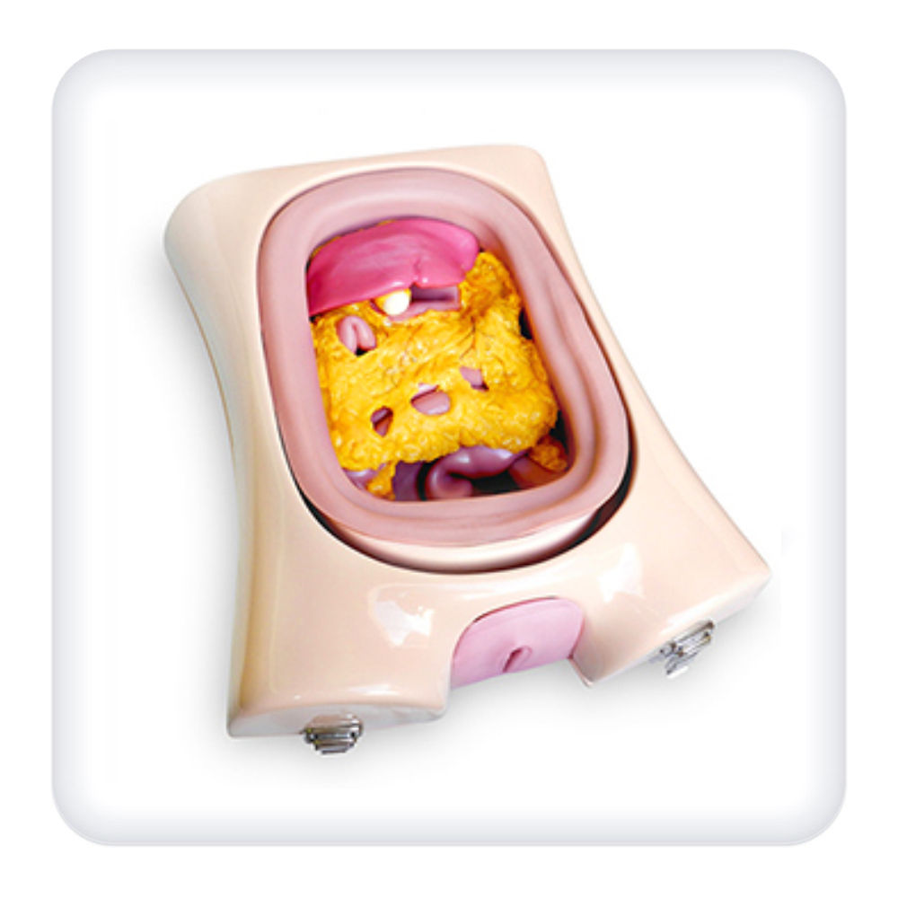
The simulator is an anatomical model of a female torso from the upper thighs to the upper chest. The front wall of the model is removable, framed and reproduces the layered anatomical structure of the anterior abdominal wall.
The simulator enables to learn the skills of endosurgical interventions on the organs of the abdominal cavity and pelvis:
- cholecystectomy;
- appendectomy;
- resection of the small and large intestine;
- myomectomy;
- polypectomy;
- removal of the uterus and appendages;
- endometriosis;
- hysteroscopy.
The simulated abdominal and pelvic cavity contains models of internal organs:
- liver;
- gall bladder;
- spleen, stomach;
- small intestine;
- large intestine;
- urinary bladder;
- ureters;
- pelvic floor muscles.
The models of internal organs are made of special silicone, which gives them natural realism.
The simulator is designed for use in medical educational institutions.
Equipment:
- Laparoscopy simulator;
- Product passport;
- User manual.


